

PDF Mercury biomagnification in the food chain of a piscivorous Biology Diagrams In the wild,

Food Chains Phylicia Hancle Biology Diagrams
Food Chains Phylicia Hancle Biology Diagrams For example lion, tiger, wolves, fox, etc. Decomposers are
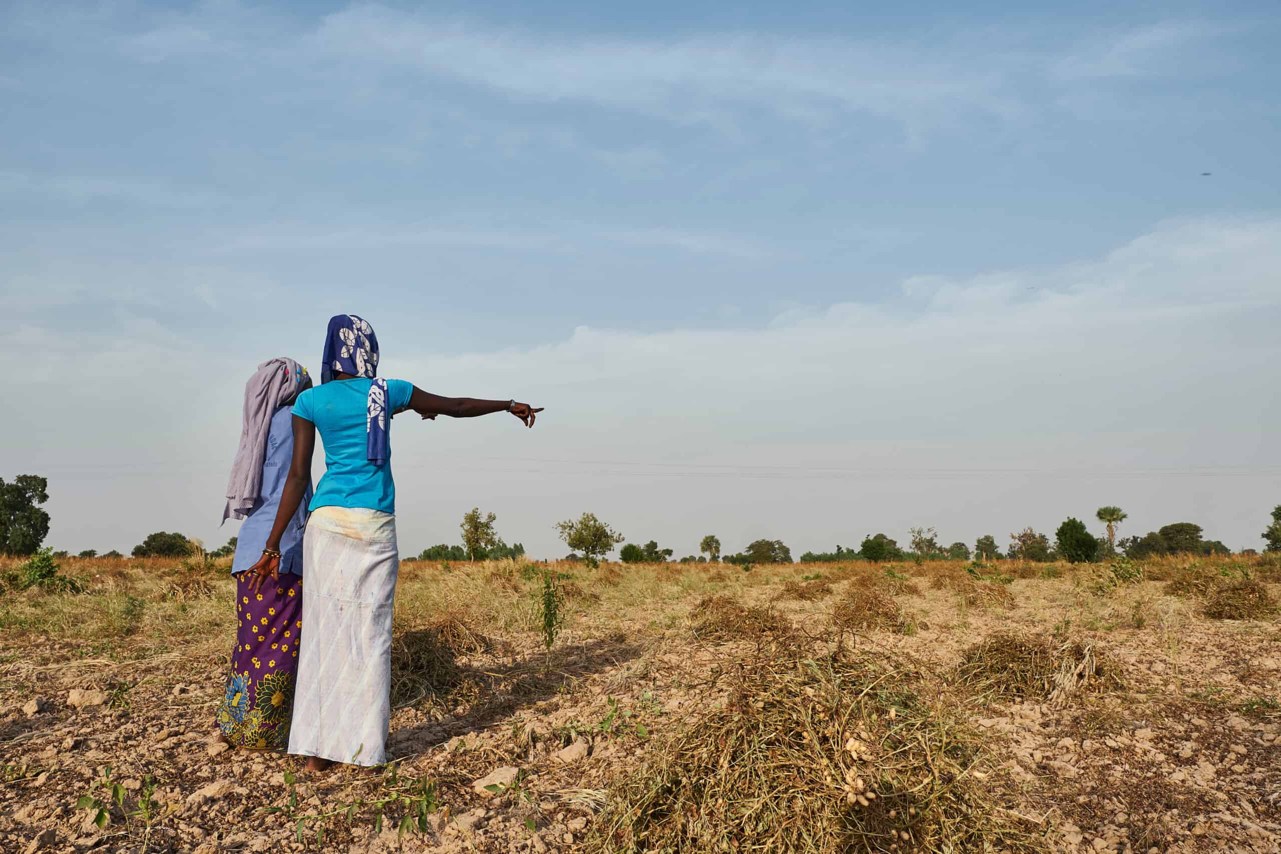
The Connection Between Food Systems and the Environment Biology Diagrams
The Connection Between Food Systems and the Environment Biology Diagrams (a) An intact food web;
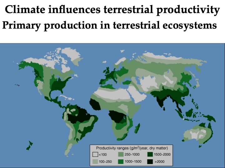
Topic 05 Primary Productivity Biology Diagrams
Topic 05 Primary Productivity Biology Diagrams Primary productivity is vital for the health of ecosystems

The nuclear envelope is constructed by the re Biology Diagrams
The nuclear envelope is constructed by the re Biology Diagrams Nuclear envelope breakdown occurs by
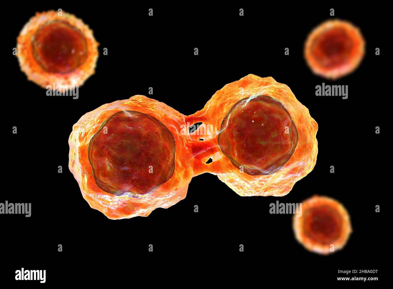
Dividing stem cells illustration Stock Photo Biology Diagrams
Dividing stem cells illustration Stock Photo Biology Diagrams The niche and asymmetric cell division of

Stem Cell Differentiation Diagram Biology Diagrams
Stem Cell Differentiation Diagram Biology Diagrams Human embryonic stem cells (hESCs) are uniquely dedicated to

Histone Modifications in Stem Cell Development and Their Clinical Biology Diagrams
Histone Modifications in Stem Cell Development and Their Clinical Biology Diagrams The histone modifications are

The Elbow Joint Biology Diagrams
The Elbow Joint Biology Diagrams The elbow joint, although non-weight bearing, may be the most
.jpg)
Chapter 1 Introduction to Human Anatomy and Physiology Biology Diagrams
Chapter 1 Introduction to Human Anatomy and Physiology Biology Diagrams Digestive system - anterior view.
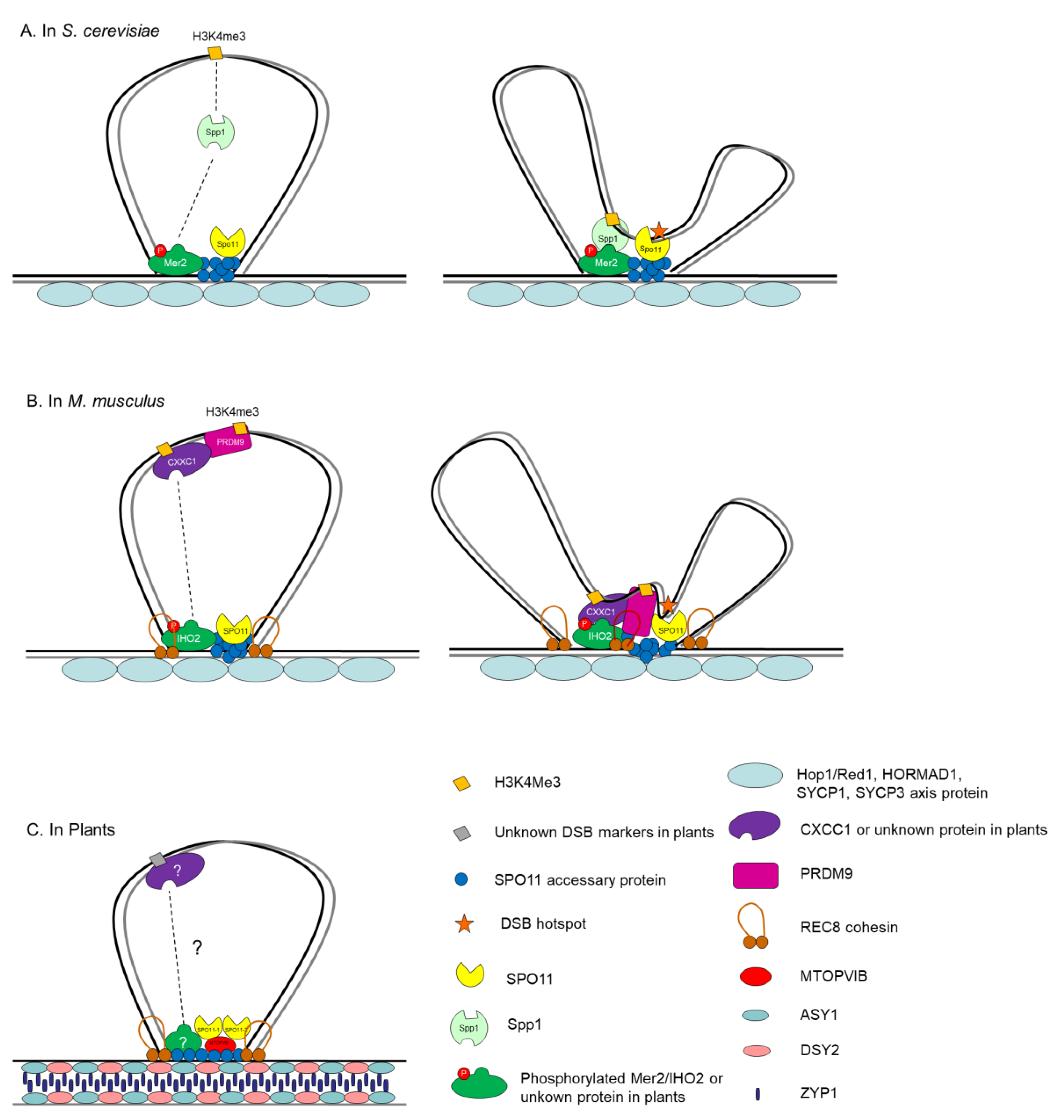
Advances Towards How Meiotic Recombination Is Biology Diagrams
Advances Towards How Meiotic Recombination Is Biology Diagrams Introduction. Meiosis is widely conserved in sexually
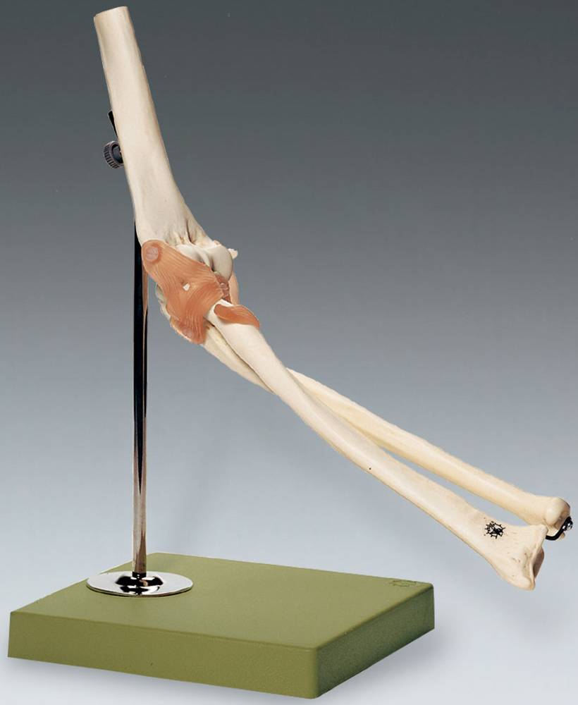
Functional Model of the Elbow Joint Anatomical Chart Company NS Biology Diagrams
Functional Model of the Elbow Joint Anatomical Chart Company NS Biology Diagrams The elbow represents
1 Draw a food chain for Puget Sound using these org Biology Diagrams
1 Draw a food chain for Puget Sound using these org Biology Diagrams The freshwater

BrdU Mouse Alexa Fluor 555 Clone 3D4 BD 100 Tests Alexa Fluor 555 Biology Diagrams
BrdU Mouse Alexa Fluor 555 Clone 3D4 BD 100 Tests Alexa Fluor 555 Biology Diagrams
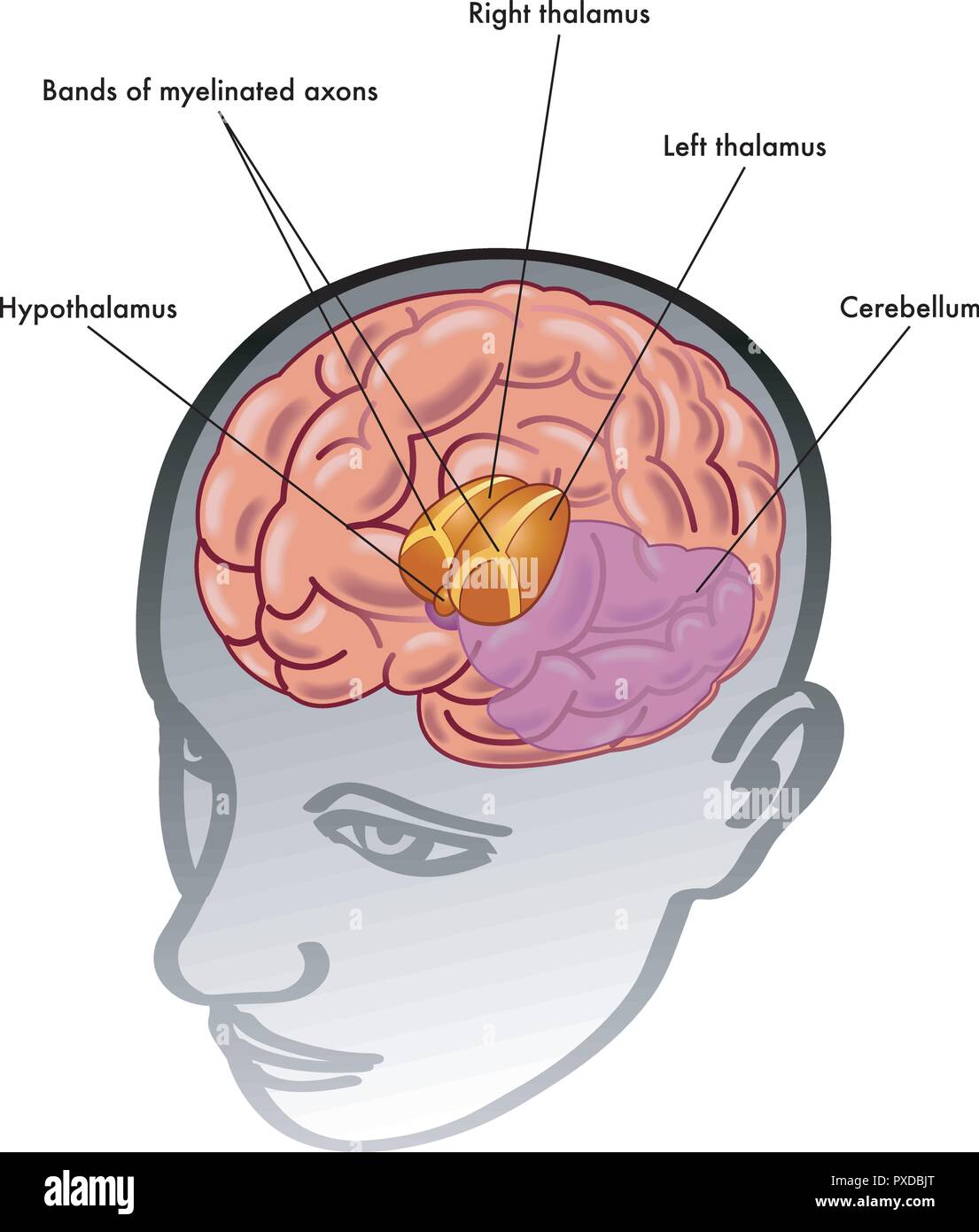
Thalamus And Hypothalamus Biology Diagrams
Thalamus And Hypothalamus Biology Diagrams The thalamus is located in the center of the brain,

What is Soil Pollution Definition Causes Effects and Solutions Biology Diagrams
What is Soil Pollution Definition Causes Effects and Solutions Biology Diagrams Pollution disrupts food chains
Anatomy radialnerve Biology Diagrams
Anatomy radialnerve Biology Diagrams The Radial Nerve (n. radialis; musculospiral nerve), the largest branch of

G1S transition Biology Diagrams
G1S transition Biology Diagrams Finally, a full accounting of how growth regulates the G1/S transition

Explain to Kids Food Chains LittleLives Biology Diagrams
Explain to Kids Food Chains LittleLives Biology Diagrams Prey-Predator Dynamics: The American house spider is
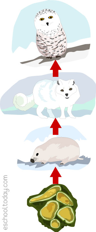
The food chain in the tundra biome Biology Diagrams
The food chain in the tundra biome Biology Diagrams A tundra food chain shows the